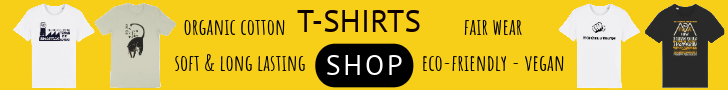
Midge Pupa
Photographer Igor Siwanowicz took these incredible photographs of tiny creatures. Dr Siwanowicz, a neurobiologist at the Howard Hughes Medical Institute’s Janelia Farm Research Campus, Virginia, shows us the wonders of life in kaleidoscopic color. Produced with a confocal laser-scanning microscope, his work enlarged on the art of Ernst Haeckel’s vivid illustrations of microscopic life, Wilson Alwyn Bentley’s stellar pictures of snowflakes, Arthur E Smith’s photo-micrographs and Daniel Kariko’s intriguing images of mini-bests in our homes.
Portrait of a Chrysopa sp. (green lacewing) larva
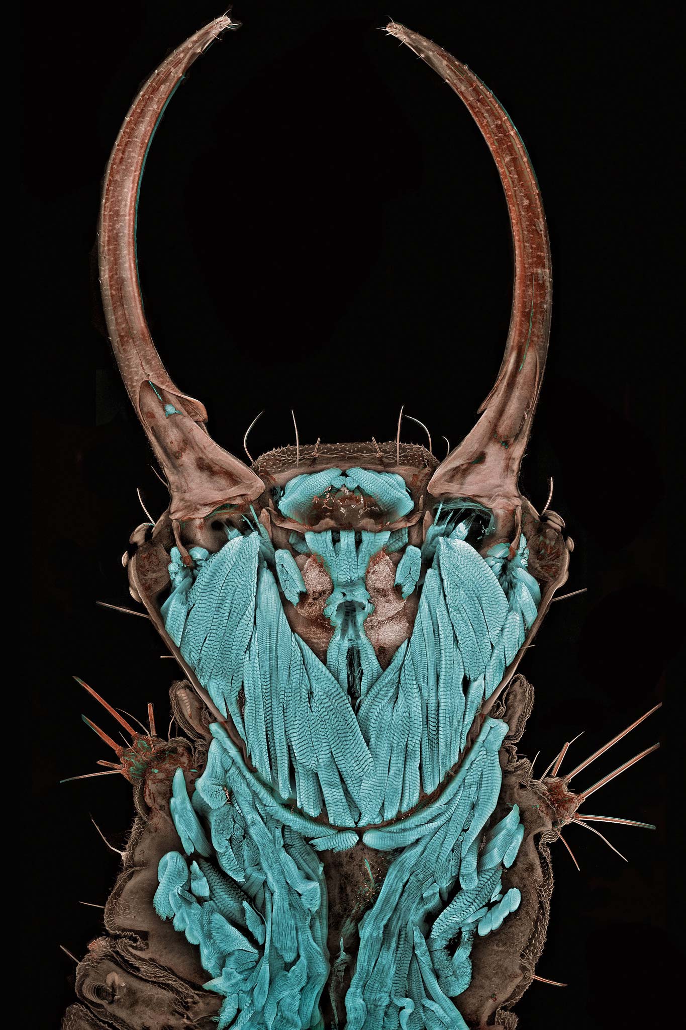
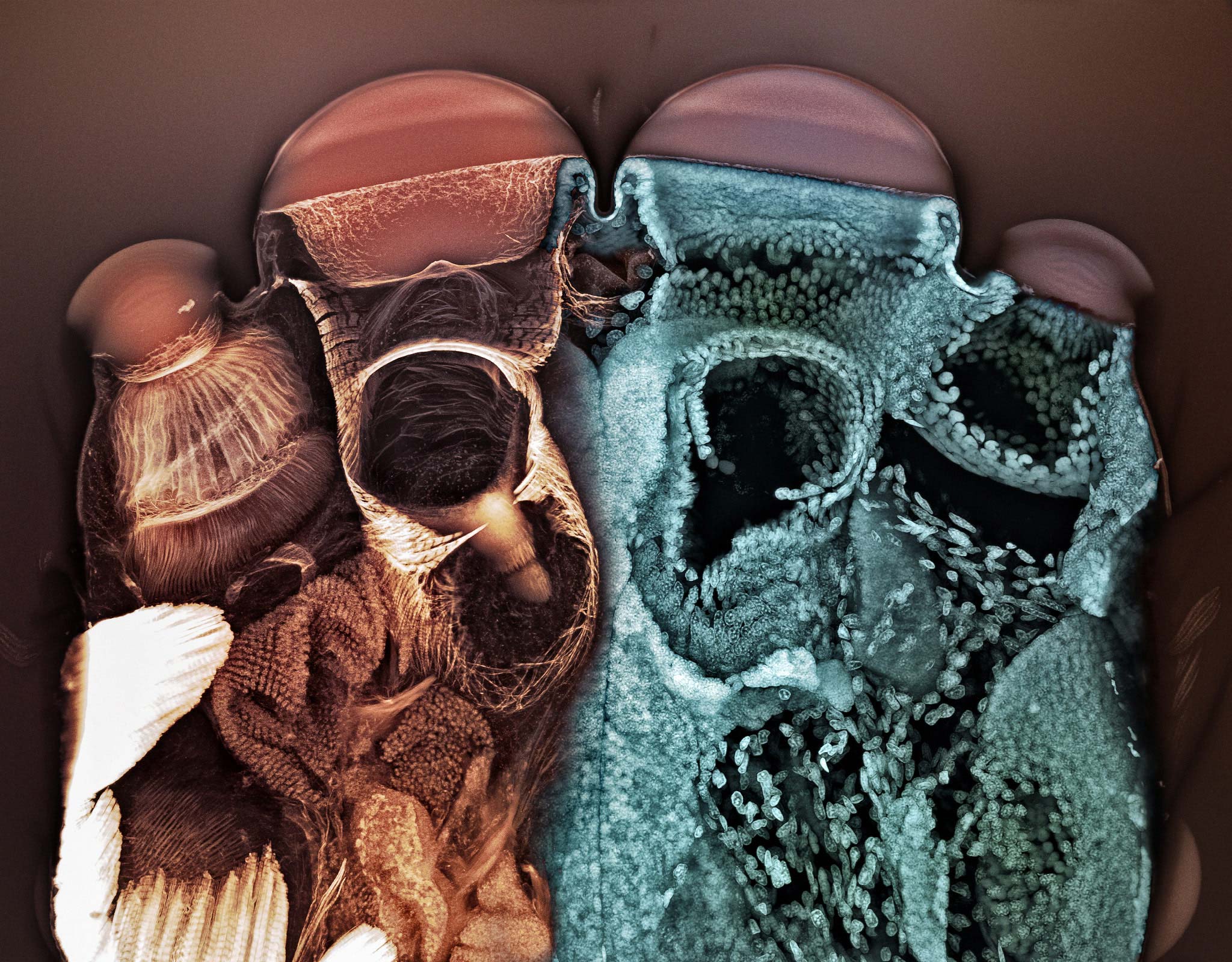
Underside of the brown dog tick and lone star tick mouthparts x 100
Confocal imaging involves scanning the specimen to create computer-generated optical sections down to 250 nm thickness using visible light. These optical sections may be stacked to provide a 3-D digital reconstruction of the specimen.
He explains:
Laser scanning confocal microscope produces images in a very different way than a bright field microscope (your standard biology class microscope). It is a fluorescent microscope, which means that the imaged specimen is illuminated with light of a certain wavelength and emits light of a different, longer wavelength. It’s the same physical phenomenon that makes black light posters from the ’70s-‘80s glow. The microscope, which registers that light, takes a series of images of the tiny specimen by scanning it point by point. Because the specimen is much thicker than the plane of focus, a series of images—called “stack”—is collected by moving the sample up or down. From those “optical slices” a three-dimensional image of the structures within the sample can be reconstructed.
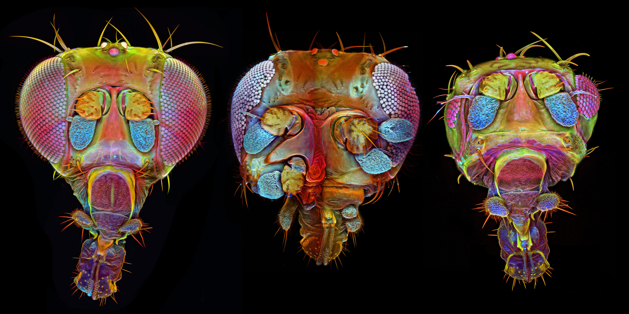
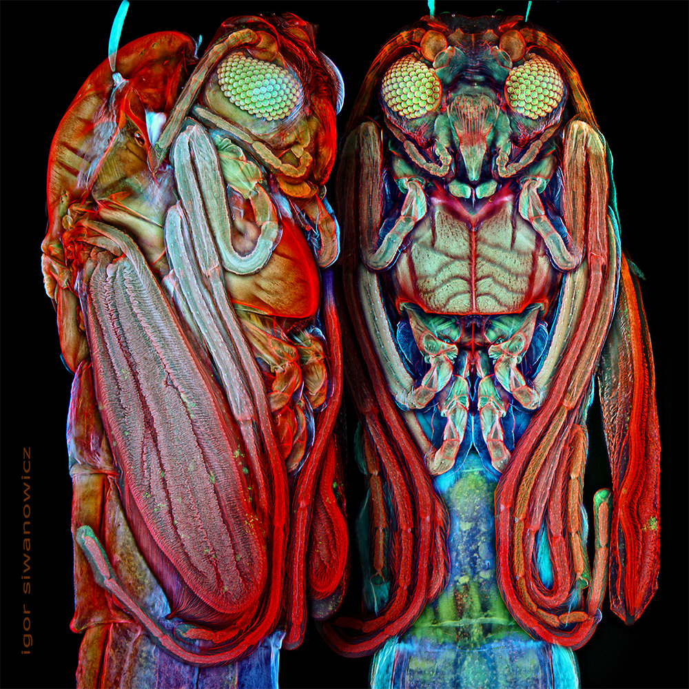
Drosophila (fruit fly) with mutant variations (middle and right images) x 250
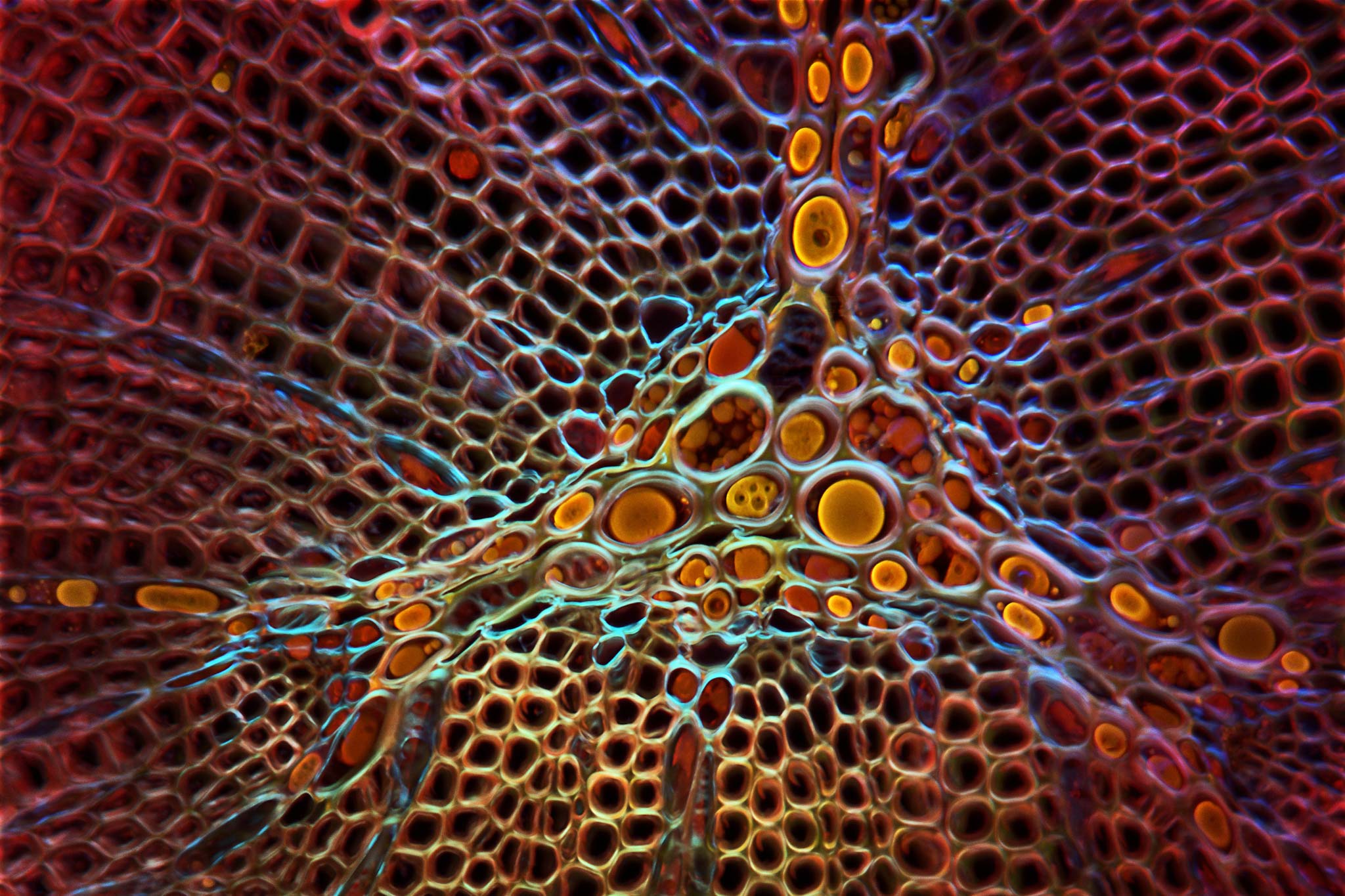
Young juniper shoot cross-section x 250
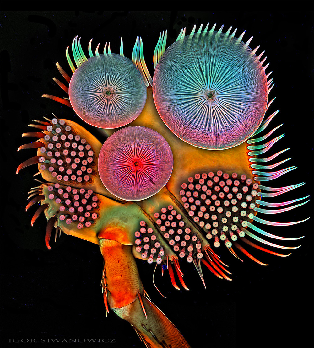
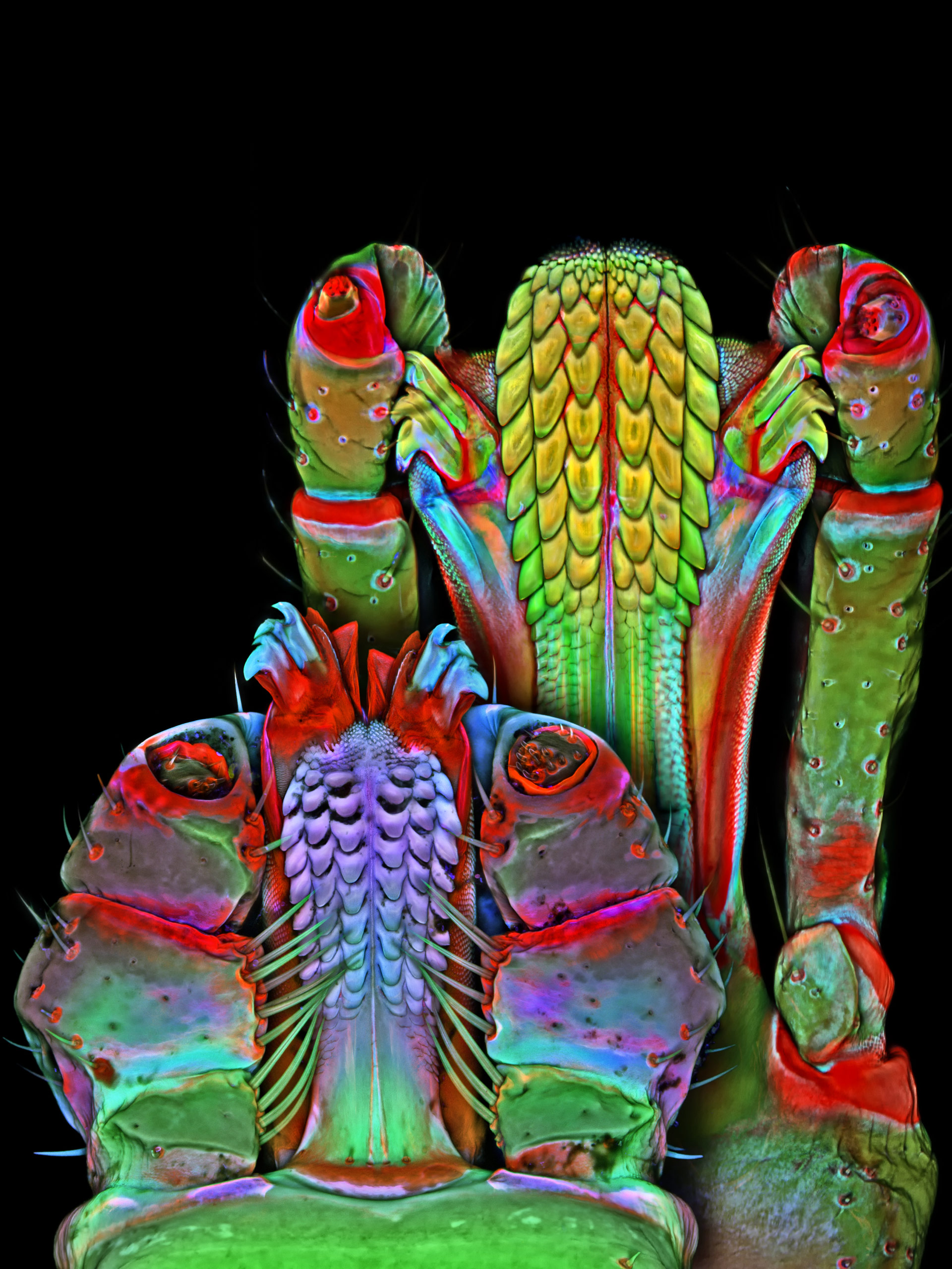
Acilius diving beetle male front tarsus (foot) 100x
Eyes of a Jumping Spider (Salticus scenicus) x 20
A Barnacle
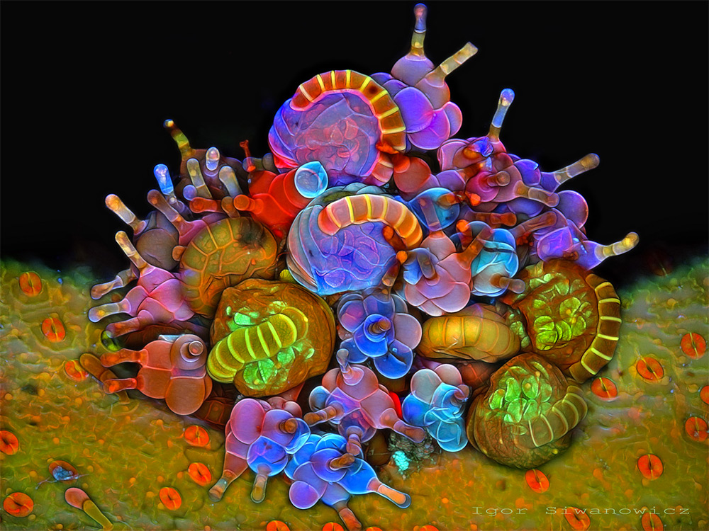
Paraphyses & Sporangia
Midge Pupa
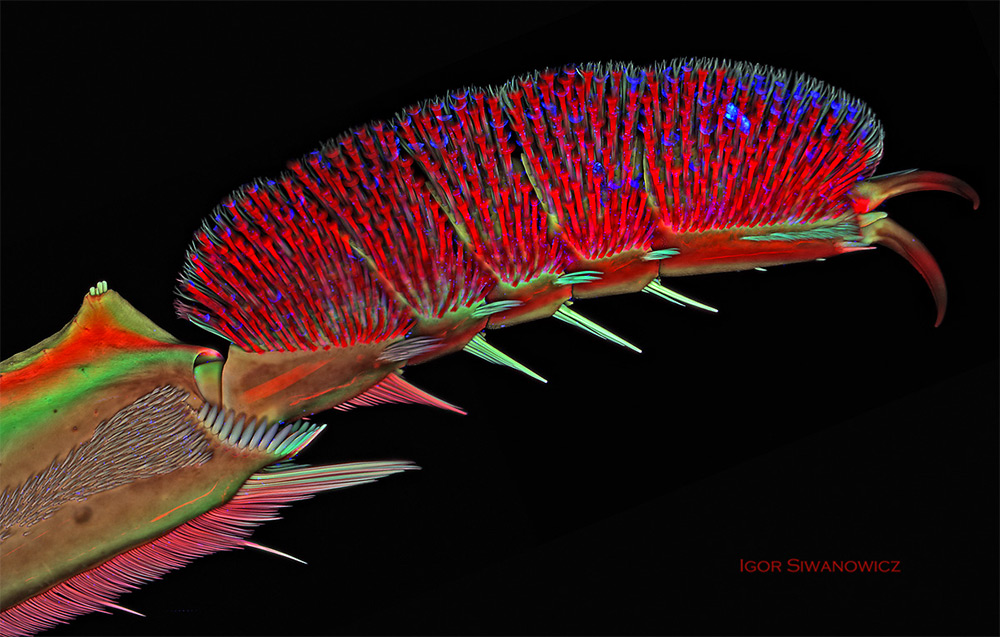
Front leg of whirligig beetle
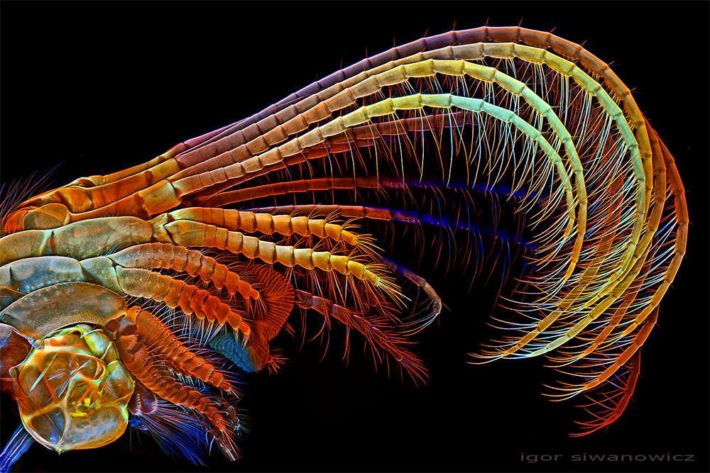
Sopod appendage
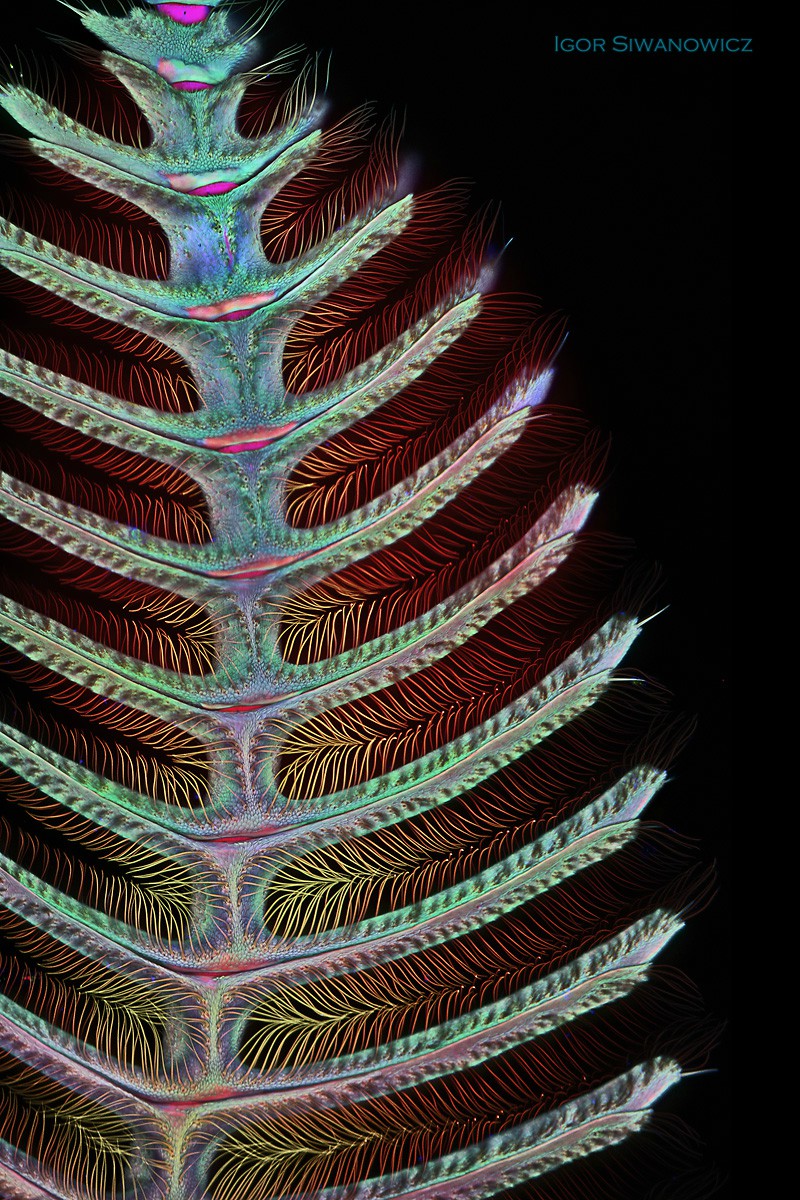
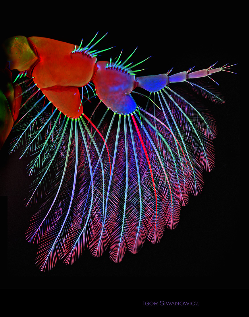
Moth antennae
Via: Nikon Small World, Wired, ThisIsColossal,
Would you like to support Flashbak?
Please consider making a donation to our site. We don't want to rely on ads to bring you the best of visual culture. You can also support us by signing up to our Mailing List. And you can also follow us on Facebook, Instagram and Twitter. For great art and culture delivered to your door, visit our shop.